The phage lambda preys upon Escherichia coli by either multiplying within its host and and destroying it (lytic cycle) or the lambda DNA can be inserted in the bacterial chromosome and remain dormant until an environmental trigger will activate this lambda (lysogenic cycle).
Selma and I investigated this lambda virus through the use of restriction enzymes, enzymes that cut DNA or they are also made to restrict the proliferation of invading viruses, in this case being the lambda virus. These are the steps to carry out the experiment:
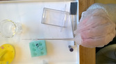
1. Using a syringe (preferably microsyringe), we put 20 micro litres of Lambda DNA into 4 different coloured wells, each containing a different restriction enzyme (one well had none, EcoRI, BamHI, Hindlll)
2. Place the wells into a holder that will float in water that has a temperature of 37 degrees. Let this sit for at least 45 minutes so the restriction enzymes have time to work properly.
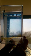
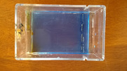
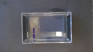
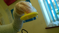
3. The agrose gel must prepared now. Molten agrose(10-12mL) must be added into an electrophoresis tank, which has to contain a comb, which leaves wells inside the gel, when removing it later. Apply the agrose gel generously.
4. place carbon fibre tissue (electrodes) on each side of the tank, so that their is created a positive and negative charge, remember the DNA is negatively charged so the DNA fragments will move towards the positive electrode. Large fragments will move slower that smaller fragments through the gel.
5. TBE buffer solution is poured creating a top layer, preventing dryness to occur.
6. Now the different enzyme solutions must be mixed with dye and loaded into the wells (which were formed by removing the comb)
7. The gel is connected to a battery, so that the DNA starts running through the gel
8. The buffer can now be poured off and a staining solution has to go on the surface of the gel for a few minutes.
9.The stain may also be removed and washed off using an ethanol solution.
10. Separate parts of the DNA should be now be seen, its easier to see on top of a white surface.
Selma and I investigated this lambda virus through the use of restriction enzymes, enzymes that cut DNA or they are also made to restrict the proliferation of invading viruses, in this case being the lambda virus. These are the steps to carry out the experiment:
1. Using a syringe (preferably microsyringe), we put 20 micro litres of Lambda DNA into 4 different coloured wells, each containing a different restriction enzyme (one well had none, EcoRI, BamHI, Hindlll)
2. Place the wells into a holder that will float in water that has a temperature of 37 degrees. Let this sit for at least 45 minutes so the restriction enzymes have time to work properly.
3. The agrose gel must prepared now. Molten agrose(10-12mL) must be added into an electrophoresis tank, which has to contain a comb, which leaves wells inside the gel, when removing it later. Apply the agrose gel generously.
4. place carbon fibre tissue (electrodes) on each side of the tank, so that their is created a positive and negative charge, remember the DNA is negatively charged so the DNA fragments will move towards the positive electrode. Large fragments will move slower that smaller fragments through the gel.
5. TBE buffer solution is poured creating a top layer, preventing dryness to occur.
6. Now the different enzyme solutions must be mixed with dye and loaded into the wells (which were formed by removing the comb)
7. The gel is connected to a battery, so that the DNA starts running through the gel
8. The buffer can now be poured off and a staining solution has to go on the surface of the gel for a few minutes.
9.The stain may also be removed and washed off using an ethanol solution.
10. Separate parts of the DNA should be now be seen, its easier to see on top of a white surface.
 Flux RSS
